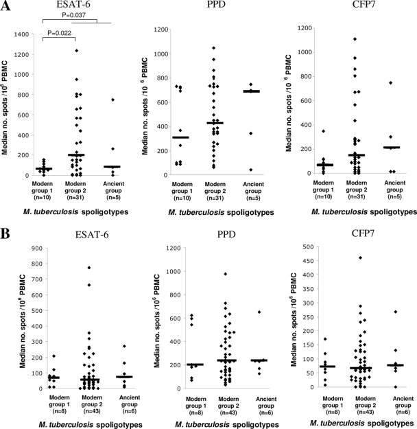
FIG. 2.
In vitro IFN-γ responses of PBMCs from ICs to restimulation with M. tuberculosis ESAT-6, PPD, and CFP7 antigens at the inclusion period (A) and after treatment (B), according to the spoligotypes of the infecting M. tuberculosis strains. The levels of IFN-γ which were significantly different between groups (Mann-Whitney test) are indicated. The horizontal bars indicate the median number of spots per 106 PBMCs.

