
FIG. 2.
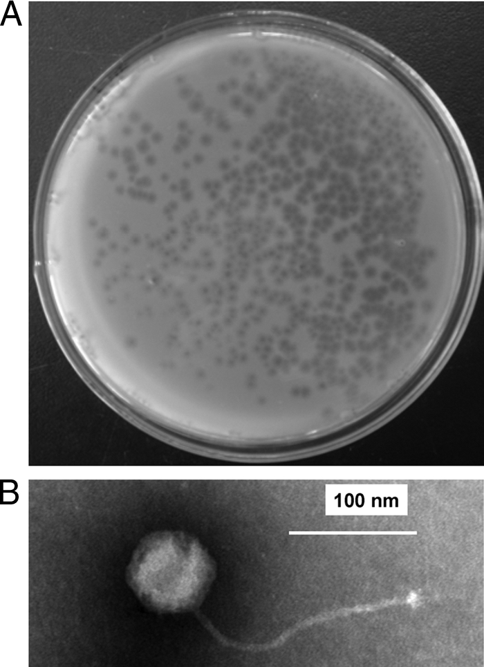
Plaques on a plate and a phage particle viewed by transmission electron microscopy. (A) Formation of plaques. Lytic φZL12 virions on a DNB plate were overlaid with soft nutrient agar containing 9R-2X spores. (B) Phage particle viewed by transmission electron microscopy. Bar, 100 nm.