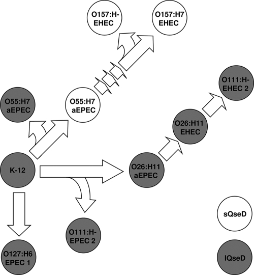
FIG. 8.
Evolution and prevalence of the various isoforms of QseD in E. coli. Shown is a cartoon representation of the evolution of EPEC and EHEC from the prototypical nonpathogenic K-12 ancestor. Solid (gray) strains represent the presence of lQseD, and open (white) strains represent the presence of sQseD (designed using original data from reference 18).

