FIG. 6.
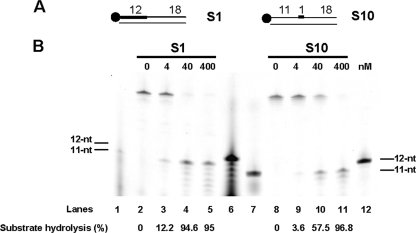
PabRNase HII specifically cuts a single embedded ribonucleotide in a DNA duplex. (A) Structures of substrates S1 and S10. The thick line and the closed circle represent the RNA portion and the fluorescent label, respectively. (B) The indicated amounts of PabRNase HII were incubated with the S1 substrate (lanes 2 to 5) and the S10 substrate (lanes 8 to 11). Both substrates and the corresponding hydrolyzed products were manually labeled. Lanes 1 and 6 are appropriate 11- and 12-nt ladders for the hydrolyzed S1 substrates. Lanes 7 and 12 are suitable 11- and 12-nt ladders for the hydrolyzed S10 substrates. Fluorescently labeled products were visualized with a Mode Imager Typhoon 9400 (GE Healthcare), and quantification was performed using ImageQuant 5.2 software.

