FIG. 6.
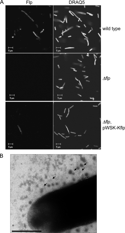
Flp is associated with the bacterial surface. (A) Confocal fluorescence microscopy indicates that Flp is expressed by and transported to the surface of the PypB-overproducing Y. enterocolitica wild type and the complemented Δflp mutant strain but not by the Δflp mutant. Flp was detected by using Flp-specific antiserum and a Cy3-conjugated secondary antibody (left), and DNA was stained with DRAQ5 (right). (B) TEM of a negative-contrasted sample indicates the presence of short pili (arrows) on the surface of the wild-type strain overproducing PypB (bar, 0.5 μm).

