FIG. 3.
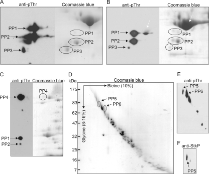
Identification of putative StkP substrates. (A) Identification of StkP substrates in the cytoplasmic fraction. Cytoplasmic proteins were separated by 2D SDS-PAGE using an IPG strip (pH 3 to 5.6) in the first dimension and a 12.5% acrylamide gel in the second dimension. The gels were stained with Coomassie blue or electroblotted and probed with anti-pThr antibody (anti-pThr). The PP1 to PP3 protein spots corresponding to phosphorylated proteins were excised and analyzed by MALDI-FTMS. (B) Identification of StkP substrates in the membrane fraction using 2D SDS-PAGE. Membrane proteins were solubilized in the presence of 4% CHAPS and separated by 2D SDS-PAGE using an IPG strip (pH 3 to 5.6) in the first dimension and a 12.5% acrylamide gel in the second dimension. The gels were stained with Coomassie blue or electroblotted and probed with anti-pThr antibody (anti-pThr). Phosphorylated proteins in the PP1, PP2, and PP3 protein spots were analyzed by MALDI-FTMS. The white arrow indicates a protein spot corresponding to enolase. (C) Identification of StkP substrates in the membrane fraction using 2D SDS-PAGE and a combination of detergents. Membrane proteins were solubilized in the presence of 4% CHAPS, 1% ASB14, 1% Triton X-100 and separated by 2D SDS-PAGE using an IPG strip (pH 3 to 10) in the first dimension and a 12.5% acrylamide gel in the second dimension. Gels stained with Coomassie blue were compared with immunoblots (anti-pThr), and the PP4 protein spot was analyzed by MALDI-FTMS. (D) Identification of StkP substrates in the membrane fraction using doubled SDS-PAGE. Membrane proteins derived from strain Sp1 were separated using a 10% acrylamide bicine-SDS-PAGE gel in the first dimension and a 8 to 16% gradient acrylamide glycine-SDS-PAGE gel in the second dimension. The gel was stained with Coomassie blue. (E) Membrane phosphoproteins separated by doubled SDS-PAGE were electroblotted, detected with anti-pThr antibody, and compared with a Coomassie blue-stained gel. Phosphorylated proteins in the PP5 and PP6 protein spots were analyzed by MALDI-FTMS. (F) Detection of StkP resolved by doubled SDS-PAGE with specific anti-StkP serum (anti-StkP). The protein corresponding to the PP5 protein spot was identified as StkP.

