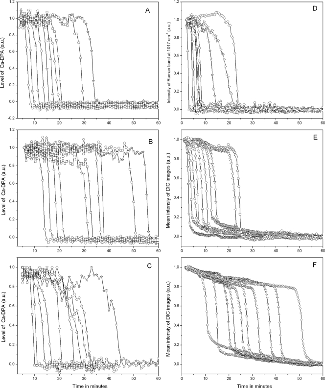
FIG. 10.
Ca-DPA levels during nutrient germination of 10 randomly chosen individual heat-activated B. subtilis spores were determined by Raman spectroscopy (A to D) and DIC microscopy (E and F). (A to C) Heat-activated (70°C) spores of B. subtilis strain PS533 (wild type) were germinated at 37°C with 10 mM L-alanine in 25 mM Tris-HCl buffer (pH 7.4) (A), 50 μM l-alanine in 25 mM Tris-HCl buffer (pH 7.4) (B), and AGFK (2.5 mM l-asparagine, 5 mg/ml d-glucose, 5 mg/ml d-fructose, 5 mM KCl) (C), and Ca-DPA levels were determined by Raman spectroscopy as described in Materials and Methods. (D and E) Ca-DPA levels in spores of strain PS3415 (200-fold elevated GerB* levels) germinated at 37°C with 5 mM l-asparagine in 25 mM HEPES buffer (pH 7.4) were monitored by Raman spectroscopy (D) and DIC microscopy (E). (F) Ca-DPA levels in spores of strain FB10 (normal GerB* levels) germinated at 37°C with 5 mM L-asparagine in 25 mM HEPES buffer (pH 7.4) were monitored by DIC microscopy.

