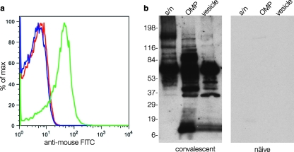
FIG. 1.
Recognition of ETEC surface antigens following experimental infection. (a) Bacterial surface recognition flow cytometry data obtained following incubation of ETEC strain H10407 with sera from mice obtained before (red peak) and after repeated enteric challenge with strain H10407 (green peak). The blue peak shows unlabeled organisms. FITC, fluorescein isothiocyanate; max, maximum. (b) Immunoblots demonstrating that multiple surface-expressed ETEC proteins are recognized by convalescent mouse sera relative to controls. Different fractions are shown (supernatant [s/n], outer membrane proteins [OMP], and outer membrane vesicles [vesicle]). The positions of molecular mass markers (in kilodaltons) are shown to the left of the immunoblot.

