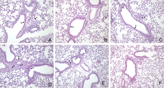
FIG. 6.
Changes in lungs of SCID mice reconstituted with CD4+ T cells, CD8+ T cells, or both CD4+ T cells and CD8+ T cells, nonreconstituted SCID mice, and BALB/c mice infected with 103 genome copies of NMI. Mice were euthanized at 24 days p.i., and lungs were fixed in formalin and stained with H&E. (A) Lung of a CD4+ T-cell-reconstituted mouse showing inflammatory cell infiltration (arrow a) and marked epithelial cell hypertrophy (arrow b). (B) Lung of a CD8+ T-cell-reconstituted mouse showing some cellular infiltration (arrow a) and minimal epithelial cell hypertrophy (arrow b). (C) Lung of a mouse reconstituted with both CD4+ T cells and CD8+ T cells exhibiting some minimal cell infiltration (arrow a) and epithelial cell hypertrophy (arrow b). (D) Lung of a SCID mouse showing extensive inflammatory changes, including marked cellular infiltration (arrow a), edema (arrow b), airway epithelial cell hypertrophy (arrow c), and bronchiolozation (arrow d). (E) Lung of an infected BALB/c mouse showing minimal cell infiltration (arrow a) and minimal changes in airway epithelial cells (arrow b). (F) Lung of an uninfected BALB/c mouse. Original magnification, ×100.

