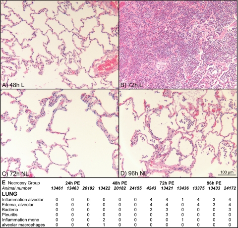
FIG. 6.
Histopathology in lung regions with gross abnormality (lesions [L]) and without gross abnormality (no lesions [NL]). (A) Normal-appearing lung with focal thickened alveolar septa (grade 1) at 48 h p.e. (B) Firm nodule (lesion) with hemorrhage and severe alveolitis (grade 4) at 72 h p.e. (C) Normal-appearing lung (no lesions) adjacent to tissue shown in panel B at 72 h p.e, with diffuse thickened alveolar septa (grade 2). (D) Normal-appearing lung (no lesions) at 96 h p.e., with congestion and alveolitis (grade 3). (E) Histopathological scores for lung tissues from 12 macaques. The “moribund” group included three animals that were euthanized because they were moribund at 70 h, 92 h, and 94 h p.e. “Inflammation alveolar” indicates predominantly polymorphonuclear leukocyte infiltration, while “inflammation mono” indicates mononuclear leukocyte infiltrate without neutrophils.

