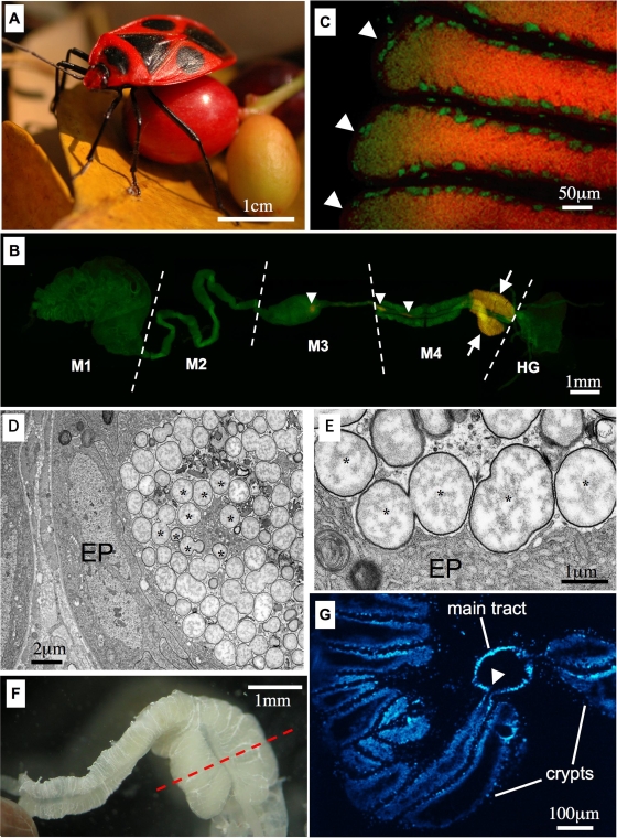
FIG. 1.
(A) Adult female of P. japonensis carrying fruit. (B and C) In situ hybridization targeting 16S rRNA of the symbiont. Red and green indicate the symbiont and the host nucleus, respectively; yellow is due to superimposed red and green signals. (B) Dissected female alimentary tract. The arrows indicate concentrated symbiont signals in the specialized symbiotic organ, swollen midgut crypts, while the arrowheads indicate sporadic symbiont signals in the midgut main tract. Abbreviations: M1, midgut first section; M2, midgut second section; M3, midgut third section; M4, midgut fourth section with crypts; HG, hindgut. (C) Enlarged image of the swollen midgut crypts. There are strong symbiont signals in the lumen of the crypts. Each arrowhead indicates a crypt. (D and E) Transmission electron microscopy of the swollen midgut crypt. Asterisks indicate symbiont cells. EP, epithelial cells of the crypt. (D) Symbiont cells restricted to the lumen of a crypt. (E) Enlarged image of symbiont cells. (F) Dissected midgut fourth section from an adult female. (G) Section of the midgut fourth section approximately through the plane indicated by the dashed line in panel F. The arrowhead indicates the connection between a crypt and the main tract of the midgut.

