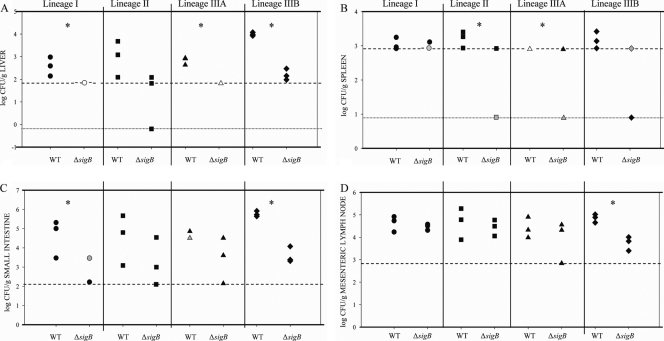
FIG. 5.
Log CFU/g L. monocytogenes recovered from organs: scatter plots of L. monocytogenes recovered from the organs of guinea pigs at 72 h after intragastric inoculation. The strains (a wild-type strain [WT] and the corresponding ΔsigB strain belonging to each lineage) are indicated on the x axis. The numbers of bacteria in the liver (A), spleen (B), small intestine (C), and mesenteric lymph nodes (D) are indicated on the y axis. Data were obtained from three guinea pigs that were intragastrically inoculated with each strain. Black symbols indicate a single data point, gray symbols indicate two overlapping data points, and open symbols indicate three overlapping data points. The detection limits, which were different for different organs due to different organ weights, are indicated by horizontal dashed and dotted lines in each panel. The dashed horizontal lines indicate the detection limit for direct plating; the dotted lines in panels A and B indicate the detection limits for enrichment procedures. The data reported for the plating detection limit were positive for L. monocytogenes after enrichment, but the bacterial counts were below the counts detectable by standard plate counting. For the data reported for the enrichment detection limit there was no recovery of L. monocytogenes after enrichment. An asterisk indicates that significantly (P < 0.05, one-sided t test) higher numbers of bacteria were recovered from organs from animals inoculated with the wild-type strain than from organs from animals inoculated with the isogenic ΔsigB mutant.

