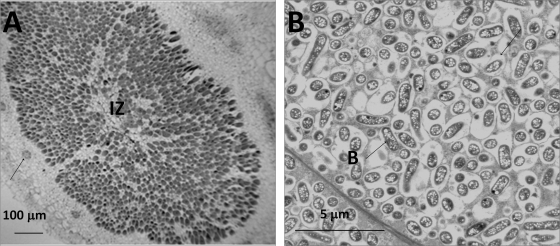
FIG. 5.
Anatomy of soybean (G. max cv. McCall) nodules formed by the USDA257 gcvT mutant. (A) Light micrograph of a paraffin section of soybean nodule revealing a central infected zone (IZ). The arrows point to vascular bundles in the cortex. (B) Transmission electron micrograph of soybean nodule showing the presence of numerous bacteroids (B) which are surrounded by symbiosomes. Note the presence of numerous poly-β-hydroxybutyrate inclusions inside the bacteroids.

