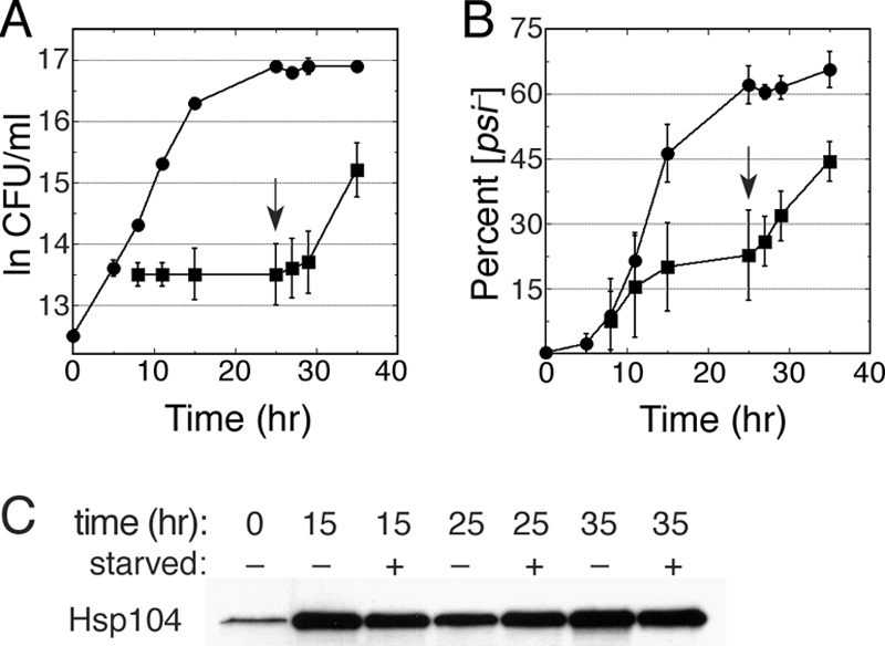
FIG. 7.

Curing of [PSI+] by Hsp104 overexpression is associated with cell division. Curing of a wild-type culture was done as described in the legend for Fig. 1 except that the culture was harvested 5 h after inducing Hsp104 expression and split into two flasks, one containing identical medium (control; circles) and the other into a similar medium lacking a required nutrient (leucine or histidine; squares). After incubation for another 20 h, the depleted nutrient was added back to the second culture (arrow). (A) Growth of cultures is shown as log of CFU per ml as a function of time. (B) The proportion of [psi−] colonies in the same culture aliquots used to monitor growth is shown. Values in panels A and B are averages from the results for three independent experiments, (two with leucine depleted, one with histidine depleted) ± standard deviation. (C) Western analysis of Hsp104 abundance in cells of the same aliquots of starved (+) and unstarved (−) cultures removed at the indicated time points from the experiment shown in panels A and B.
