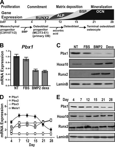
FIG. 1.
Pbx1 expression decreases during osteoblast differentiation. (A) Schematic illustrating temporal gene expression during osteoblast differentiation. Osteoblast-like differentiation was induced in C3H10T1/2 cells by treatment with either 2% FBS (control), 100 ng/ml BMP2, or 10 nM dexamethasone for 7 days. (B) Relative mRNA expression of Pbx1 in C3H10T1/2 cells was determined by RT-qPCR. (C) Protein expression was determined by Western blotting using an anti-Pbx1 antibody. MC3T-3E1 cells were treated with 280 μM ascorbic acid-5 mM β-glycerol phosphate to induce osteogenic differentiation for a total period of 28 days. (D) Total RNA was isolated, and relative expression of TALE protein family genes was determined by real-time quantitative PCR using gene-specific primers. (E) Relative protein expression of Pbx1 over the 28-day time course was determined by Western blotting using an anti-Pbx1 antibody. Error bars indicate SEM.

