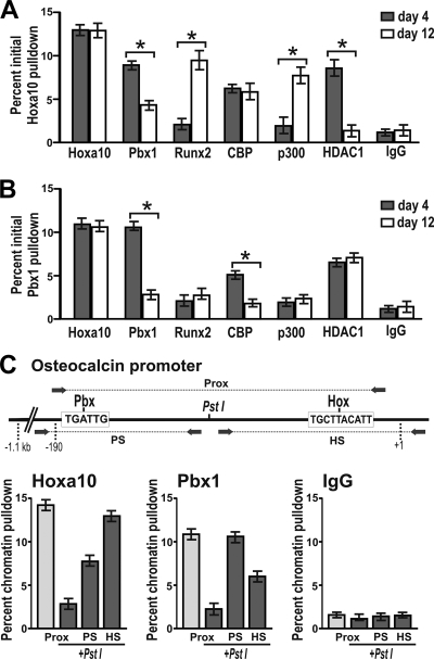
FIG. 7.
Pbx1 cooccupancy with repressive histone-modifying enzymes and Hoxa10 on the osteocalcin promoter in proliferating cells. Cross-linked cell lysates from primary calvarial osteoblasts were immunoprecipitated using either anti-Hoxa10 or anti-Pbx1 antibody, and the resulting fraction was further immunoprecipitated using the specified antibodies (ChIP-reChIP). (A) The Hoxa10-immunoprecipitated fraction was further immunoprecipitated using Hoxa10, Runx2, Pbx1, CBP, p300, HDAC1, or control (IgG) antibodies and quantified by qPCR. (B) The Pbx1-immunoprecipitated fraction was further immunoprecipitated using Hoxa10, Runx2, Pbx1, CBP, p300, HDAC1, or control (IgG) antibodies and quantified by qPCR. (C) Cross-linked chromatin from proliferating calvarial osteoblasts was treated with PstI to cleave DNA between the Hoxa10- and Pbx1-binding sites, immunoprecipitated with anti-Hoxa10 or anti-Pbx1 antibody, and evaluated with primer sets specific for digested (Pbx site [PS] or Hox site [HS]) or undigested (proximal promoter [Prox]) fragments. Statistical significance was determined by Student's t test (*, P < 0.05 versus matched control). the ChIP experiment was repeated three times with similar results, and the data presented are from one representative experiment (± SD).

