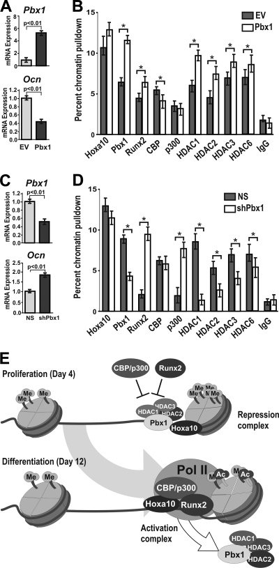
FIG. 9.
Modification of Pbx1 levels in osteoblasts results in alteration of histone-modifying enzymes at the osteocalcin promoter. (A and B) Rat calvarial osteoblasts were isolated from embryonic day 18.5 rat pups and infected with Pbx1 or empty (EV) lentiviral constructs, and cells were collected at 48 h after infection after just reaching confluence (day 6). (A) Relative expression of the Pbx1 and Ocn genes was monitored by RT-qPCR. (B) ChIP analysis was performed on cleared lysates from primary osteoblasts using ∼5 μg of the indicated antibody. Recovered DNA was then quantified by qPCR using primers specific for the proximal promoter region of the osteocalcin gene to determine the relative occupancy of the indicated proteins. (C) Isolated osteoblasts (as described above) were infected with Pbx1-shRNA or nonsilencing shRNA (NS) lentiviral constructs, and relative expression of Pbx1 and Ocn was determined by RT-qPCR. (D) ChIP analysis was performed on cleared lysates from treated primary osteoblasts (as described above) to determine the relative occupancy of the indicated proteins on the osteocalcin promoter. Statistical significance was de- termined by Student's t test (*, P < 0.05 versus matched control). ChIP experiments were repeated two times with similar results, and the data presented are representative of one experiment (± SD). (E) Schematic model of Pbx-mediated repression of the osteocalcin gene.

