FIG. 4.
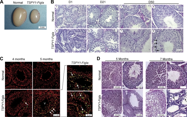
Testicular dysmorphology of TSPY1-Figla mice. (A) At 7 weeks, the TSPY1-Figla testes were smaller than normal and the average weight (mean ± SEM) was 73.6 ± 3.1 mg, compared to 100 ± 2.1 mg for normal mice. (B) Cross-sections of seminiferous tubules of normal and transgenic mice at D1, D21, and D50 after fixing in Bouin's solution and staining with hematoxylin and counterstaining with eosin. Arrows (panel 8) indicate enlarged spermatocytes. (C) TUNEL assay at 4 and 5 months of normal and TSPY1-Figla transgenic testes. Apoptotic cells, Hoechst 33242-stained nuclei, and merged images are green, red, and yellow, respectively. Roman numerals refer to the stages of mouse spermatogenesis. Arrows indicate apoptotic germ cells. (D) Testicular histology (fixed in Bouin's solution and stained with periodic acid-Schiff's reagent and eosin) of normal and TSPY1-Figla transgenic animals at 5 and 7 months. Abnormal spermatogenesis and vacuolation (arrows) that were accentuated by age were observed in transgenic tubules.

