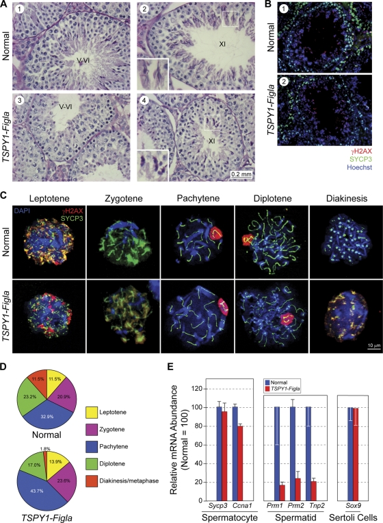
FIG. 5.
Meiosis in TSPY1-Figla male mice. (A) Testicular histology of normal and TSPY1-Figla transgenic mice at 5 months. Few postmeiotic cells were present in stage V and VI tubules from transgenic mice (panels 1 and 3), and those in stage XI had foreshortened heads (panels 2 and 4, insets). Stages in the seminiferous epithelial cycle are indicated by Roman numerals. (B) Testicular sections from 4-month-old normal and TSPY1-Figla transgenic mouse testicular tubules were stained with antibodies to SYCP3 (green), a synaptonemal protein, and γH2AX (red), a phosphorylated histone variant, indicative of double-strand breaks and regions of asynapsis. Both proteins were detected in peripherally located spermatocytes, and γH2AX was also detected in the more centrally located elongating spermatids. Nuclei were stained with Hoechst 33242. (C) Immunofluorescence of chromatin spreads from mixed germ cells isolated from 4-month-old normal or TSPY1-Figla transgenic mouse testes and probed with antibodies to SYCP3 (green) and γH2AX (red). Nuclei were detected with Hoechst 33242. (D) Pie graphs indicate the percentage of spermatocytes in each of the substages of the prophase of the first meiotic division of normal mice (680 cells) and TSPY1-Figla transgenic mice (798 cells). (E) qRT-PCR analysis (mean ± SEM) of expression levels of selected genes in spermatocytes, spermatids, and Sertoli cells from 5-month-old normal and TSPY1-Figla transgenic mouse testes.

