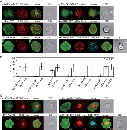
FIG. 1.
Images of lipid raft-associated proteins and lipid raft probes at TCR-MCs. (A) AND-Tg T cells expressing mLAT(wt)-GFP, mLAT(ΔCP)-GFP, Lck-GFP, or Lck(N10)-GFP together with CD3ζ-Halo, or cells expressing CD3ζ-Halo and stained with CTXB-Alexa 488, were loaded with TMR ligand and placed on the planar bilayer containing ICAM-I and I-Ek with 10 μM MCC peptide. Images were collected 2 min after attachment to the bilayer. The cells expressing Lck(N10)-GFP and CD28-CFP were fixed at 2 min on the CD80-containing bilayer and stained with anti-CD3ɛ-biotin and streptavidin-Alexa 566. The large bright area in cells with mLAT(ΔCP)-GFP reflects its presence within cytoplasmic organelles as detected in the x-z image (see Fig. S1 in the supplemental material). (B) The numbers of GFP and Halo tag clusters were counted objectively using software with Gaussian fitting and counting capability. Data are means ± standard deviations of 15 to 30 cells. (C) The cells used in panel A were incubated for 10 min, and images were collected. DIC, differential interference contrast.

