FIG. 5.
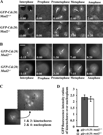
Cdc20 kinetochore recruitment and localization is Mad2 independent. (A and B) A gfp-cdc20 transgene on the second chromosome was introduced into the mad2 wild-type (w67) and the mad2EY-null mutant lines, and time-lapse images were acquired for comparison: confocal images show GFP-Cdc20 in syncytial embryos (A), and DeltaVision fluorescence microscope images show GFP-Cdc20 in third-instar larval neuroblasts (B). (Top rows) Wild-type embryos. (Bottom rows) mad2EY-null (mad2−/−) mutant embryos. The arrowheads indicate the exclusion of GFP-Cdc20 signal from the interphase nucleus. The arrows indicate kinetochore-associated GFP-Cdc20. Bar = 5 mm. (C) Sample image showing a selection of the region of interest on individual kinetochores or nearby nucleoplasm regions for fluorescence intensity quantification and ratio analysis of the GFP-Cdc20 kinetochore signal at its peak time. (D) Quantitative comparison of GFP-Cdc20 kinetochore/nucleoplasm ratio signals between the wild type and mad2EY-null mutant; 16 kinetochores from 4 different embryos or neuroblasts were analyzed. The error bars indicate standard deviations.

