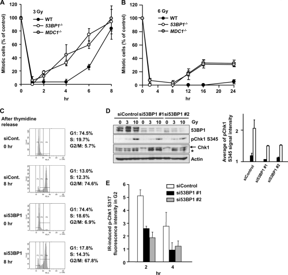
FIG. 5.
MDC1−/− and 53BP1−/− MEFs show impaired G2/M checkpoint maintenance and reduced Chk1 phosphorylation. (A and B) Mitotic entry in WT, MDC1−/−, and 53BP1−/− MEFs after 3 Gy and 6 Gy IR was evaluated. APH was added after IR. (C) Cell cycle profile in control and 53BP1 siRNA cells after double thymidine block. (D) p-Chk1 is reduced in synchronized G2 53BP1 siRNA cells post-IR. Cells were synchronized in G1/S by double thymidine block and irradiated 8 h postrelease. Cells were examined by immunoblotting using anti-pChk1 Ser345 antibody. The asterisk represents a nonspecific band. Quantification of p-Chk1 from 2 experiments is shown. (E) 53BP1 siRNA cells show impaired Chk1 activation. The p-Ser317 Chk1 signal was quantified by IF in CENP-F+ (G2) A549 cells treated 3 Gy IR and 53BP1 or control siRNA. The signal in undamaged nuclei is subtracted. Error bars represent the SEM of 3 experiments.

