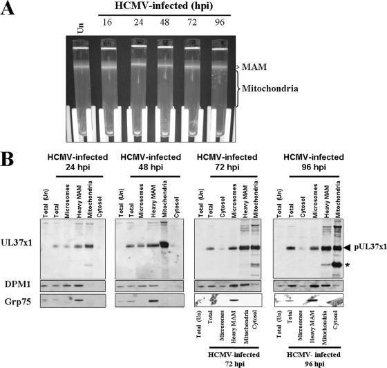
FIG. 1.
(A) HCMV infection alters mitochondrial banding in self-generated Percoll gradients. LE-HFFs were infected with HCMV (AD169, 3 PFU/cell). Four roller bottles of infected cells were harvested at 16, 24, 48, 72, and 96 hpi and fractionated in each gradient as previously described (7, 8). The positions of the MAM and mitochondrial fractions are indicated on the gradients. (B) pUL37x1 is detected in the heavy MAM fraction of HCMV-infected LE-HFFs. The microsomal, heavy MAM (6,300 × g), mitochondrial, and cytosolic fractions from uninfected cells (Un) and HCMV-infected cells were obtained from the fractionations shown in panel A at 24, 48, 72, and 96 hpi. Ten-microgram amounts of total and fractionated proteins were separated by SDS-PAGE and examined by Western blot analyses using rabbit anti-UL37x1 antiserum (DC35, 1:1,000). The fractions were reacted with antibody against an ER marker (DPM1, 1:100) or a mitochondrion/MAM marker (Grp75, 1:1,000). The order of samples used for the Grp75 blots for HCMV-infected LE-HFFs at 72 and 96 hpi is indicated below the corresponding panels. The positions of pUL37x1 (arrowhead) and of a short UL37 species (asterisk) are indicated on the UL37x1 blots.

