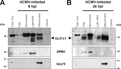
FIG. 2.
pUL37x1 is detected in the highly purified light MAM fraction from HCMV-infected LE-HFFs at IE and early times of infection. LE-HFFs were infected with HCMV (AD169, 3 PFU/cell) and were harvested at 8 (A) or 24 (B) hpi. Fractions from 4 roller bottles each of uninfected or infected cells were obtained as described for Fig. 1 and previously described (7, 8). Twenty-microgram amounts of fractionated proteins (microsomes, light MAM fraction [100,000 × g], mitochondria, and cytosol) from cells harvested at the indicated times were resolved by SDS-PAGE and examined by Western analysis using anti-UL37x1 antiserum (DC35, 1:250), as well as antibody against an ER marker (DPM1, 1:100) or a mitochondrion/MAM marker (Grp75, 1:500). The position of pUL37x1 (arrowheads) is indicated on the UL37x1 blots.

