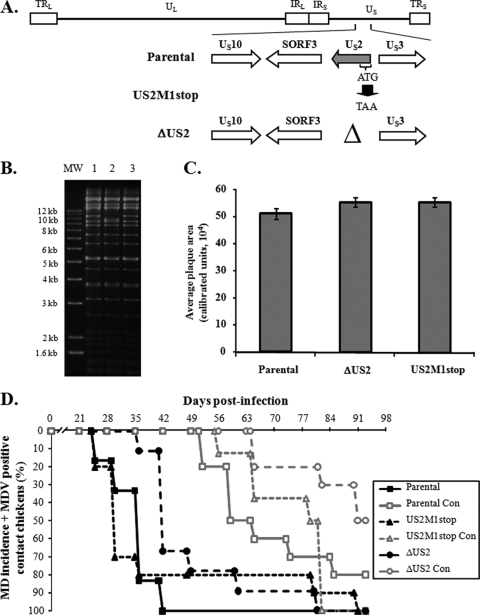
FIG. 1.
Generation of US2 mutant MDVs and evaluation of their ability to induce MD and horizontally transmit to contact chickens. (A) Two US2 mutant viruses were generated, one in which the complete US2 ORF was deleted (ΔUS2) and another where the ATG start codon was mutated to a TAA stop codon (US2M1stop). Also shown are genes flanking the US2 ORF in the US region of the MDV genome. (B) RFLP analysis of DNA obtained from parental virus (lane 1) and ΔUS2 (lane 2) and US2M1stop (lane 3) BAC clones using BamHI restriction patterns. Deletion of US2 reduces the size of the 10,354-bp fragment of the parental virus (lane 1) to 9,544 bp (lane 2). No extraneous alterations are evident in both clones. The molecular size marker (MW) used is the 1-kb Plus DNA ladder from Invitrogen, Inc. (Carlsbad, CA). (C) The average plaque area ± standard error of the mean (SEM) for each respective virus was determined from 75 plaques exactly as previously described (18). No significant differences were seen between viruses using Student's t tests. (D) MD incidence of P2a chickens inoculated at 1 day of age with reconstituted BAC clones described in the text and contact (Con) chickens housed with experimentally infected chickens over the course of 13 weeks of infection. MD incidence was determined by identification of gross lesions in dead or euthanized chickens. Chickens not succumbing to MD over the course of the experiment were terminated at 92 days p.i. Blood was collected from all remaining birds and tested for MDV genomic copies using qPCR exactly as previously described (17). For determination of horizontal transmission, contact chickens positive for MDV genomic copies in the blood were included, since the presence of MDV genomes indicated spread.

