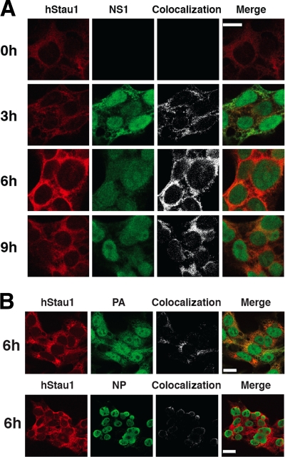
FIG. 1.
Colocalization of hStau1 protein with influenza viral proteins in infected cells. Cultures of HEK293T cells were infected with WSN virus and fixed at the times after infection indicated. The cells were processed for immunofluorescence with antibodies specific for the hStau1, NS1, NP, or PA proteins as indicated in Materials and Methods. The left columns of panels show the localization pattern of each protein. The right columns of panels show the colocalization sites of hStau1 and the viral markers used, as well as the merged images. (A) hStau1 protein is shown in red, and NS1 protein in green. The scale bar represents 10 μm. (B) hStau1 is shown in red, and either PA or NP is shown in green. The scale bar represents 20 μm.

