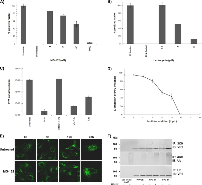
FIG. 5.
Proteasome involvement. (A and B) Inhibition of the proteolytic activity of the proteasome aborted PPV infection. The effects of the inhibitors were evaluated by indirect IF experiments as described for Fig. 2 and were expressed as the percentage of infected cells at 20 h p.i. relative to that for mock-treated cells (arbitrarily set at 100%). (C) Cells were treated with inhibitors as for panels A and B, and inhibition of genome replication was measured by qPCR for PPV, normalized to cell numbers, as described for Fig. 3. (D) Pulse inhibition. MG-132 was added at different times throughout the infection until cell fixation, and the percentage of infected cells was determined. (E) Capsid localization in the presence of MG-132. Capsids were visualized at different times throughout the infection by the capsid-specific antibody 3C9. In untreated cells, PPV reached perinuclear localization within 4 h p.i., and new virus was observed in the nucleus at 20 h p.i. The virus was also observed near the nucleus after MG-132-mediated inhibition but remained more diffuse in the cytoplasm (8 to 12 h p.i.), and very few positive nuclei were visible at 20 h p.i. (F) Coimmunoprecipitation of ubiquitin with anti-PPV capsid antibodies and of VP2 capsid proteins with anti-ubiquitin antibodies. Capsid proteins were observed in all ubiquitin precipitations. The ubiquitin signal in the capsid immunoprecipitation was more diffuse, as generally observed for ubiquitinated proteins.

