FIG. 5.
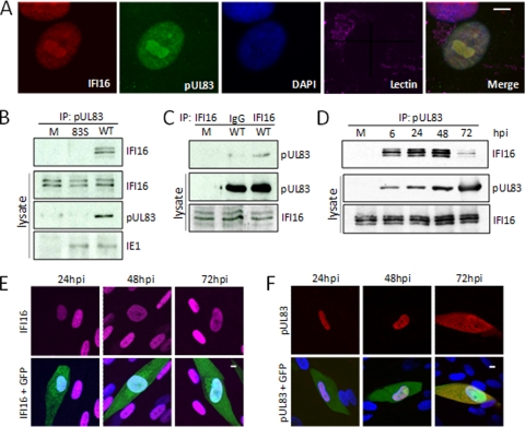
Confirmation of the pUL83-IFI16 interaction. (A) IFI16 and pUL83 were partially colocalized within infected cells. Fibroblasts infected with wild-type HCMV at a multiplicity of 0.5 PFU/cell were fixed and stained with antibodies specific for UL83 and IFI16 to test for colocalization. The nucleus and Golgi were monitored to provide context, and the white bar indicates 10 μm. (B) IFI16 coprecipitates with antibody to pUL83. Fibroblasts were mock infected (M) or infected with wild-type HCMV (WT) at a multiplicity of 3 PFU/cell and harvested 72 h later. pUL83-specific immune complexes were isolated by immunoprecipitation (IP), and the presence of IFI16 in those complexes was determined by Western blotting using antibodies specific to IFI16. Lysates were assayed by Western blotting for the presence of the indicated proteins as controls. (C) pUL83 coprecipitates with antibody to IFI16. IFI16-specific immune complexes were isolated by immunoprecipitation from mock-infected fibroblasts or at 72 hpi of fibroblasts with wild-type HCMV at a multiplicity of 3 PFU/cell. Immunoprecipitation was also performed with nonspecific antibody (IgG). The presence of pUL83 in precipitates was confirmed by Western blotting with a pUL83-specific antibody. Lysates were assayed by Western blotting for the presence of the indicated proteins as controls. (D) IFI16 interacts with pUL83 throughout the course of infection. Lysates of cells infected at a multiplicity of 3 PFU/cell were prepared at the indicated times and subjected to immunoprecipitation with antibody to pUL83. Lysates were assayed by Western blotting for the presence of the indicated proteins as controls. (E) IFI16 remained in the nucleus during HCMV infection. Fibroblasts were infected at a multiplicity of 0.5 PFU/cell with a derivative of wild-type HCMV expressing a GFP marker protein (green). Cells were fixed and processed for immunofluorescence using an antibody to IFI16 (red) at the indicated times after infection. Nuclei were stained with DAPI to provide context, and the white bar indicates 10 μm. (F) pUL83 was initially localized to the nucleus but was also in the cytoplasm by 72 hpi. Fibroblasts were infected at a multiplicity of 0.5 PFU/cell with a derivative of wild-type HCMV expressing a GFP marker protein (green). Cells were fixed and processed for immunofluorescence using an antibody to pUL83 (red) at the indicated times after infection. Nuclei were stained with DAPI to provide context, and the white bar indicates 10 μm.

