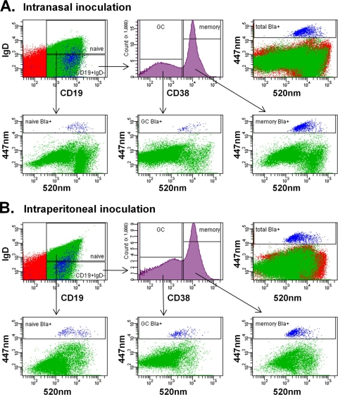
FIG. 5.
Detection of mLANA expression in mature B-cell subsets. Spleens from MHV68.ORF73βla-infected mice were harvested 16 days after intranasal (A) or intraperitoneal (B) inoculation. Single-cell suspensions were stained with antibodies to cell surface markers and then loaded with CCF2/AM β-lactamase substrate and subjected to flow-cytometric analysis. Naïve follicular B cells (CD19+ IgD+ CD38+), germinal-center B cells (CD19+ IgD− CD38low), and memory B cells (CD19+ IgD− CD38high) are depicted in the bottom panels, with mLANA/βla-positive cells (blue) indicated by the boxed gate. Data are representative of five individual experiments.

