FIG. 3.
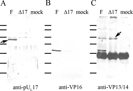
Coimmunoprecipitation of proteins with anti-VP13/14 antibody. Cells were mock infected or were infected with HSV-1(F) or the UL17 deletion virus, and lysates were reacted with VP13/VP14-specific antibody. Immune complexes were purified, electrophoretically separated, and subjected to immunoblotting with antibodies against pUL17 (A), VP16 (B), or VP13/14 (C). Bound immunoglobulins were revealed by reaction with appropriately conjugated anti-immunogobulins followed by chemiluminescent exposure to X-ray film. The arrow in panel A indicates a band containing pUL17. The arrow in panel C refers to the band containing VP13/14. The heavy band below this is the heavy chain of rabbit IgG, which reacts with the conjugated secondary antibody. The lines to the left of each panel refer to the positions of size standards. From top to bottom, they are Mrs 180,000, 130,000, 100,000, 72,000, 55,000, 40,000, and 33,000.

