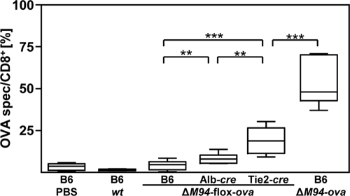
FIG. 4.
EC contribute to antiviral CD8+ T-cell stimulation. One day prior to i.p. injection of 105 TCID50 of MCMV-flox-ova-ΔM94 (ΔM94-flox-ova), MCMV-ova-ΔM94 (ΔM94-ova), MCMV-wt (wt), or PBS, 3 × 10⁵ congenic OT-I CD8+ T cells were transferred i.v. into B6, Alb-cre, and Tie2-cre mice. At day 6 p.i. a flow cytometric analysis was performed on peripheral blood lymphocytes (PBL) for the congenic marker CD45.1 and CD8. Boxes represent the ratios of OT-I cells per CD8+ cells as a pool of 3 independent experiments and extend from the 25th to the 75th percentile. The lines indicate the medians. Whiskers extend to show the extreme values. The P values were obtained by applying a two-tailed Wilcoxon rank sum test (**, P < 0.01; ***, P < 0.001).

