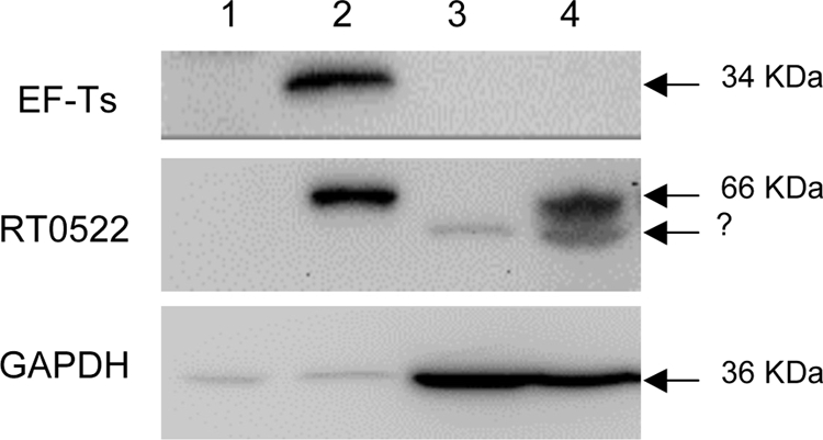
FIG. 8.

Translocation assay of RT0522 and EF-Ts by Western blotting. The membrane was probed with rabbit anti-EF-Ts antibody, rabbit anti-RT0522 antibody, or mouse anti-GAPDH monoclonal antibody by using the SuperSignal West Pico chemiluminescent substrate kit. Lane 1, pellet of uninfected Vero76 cells; lane 2, pellet of R. typhi-infected Vero76 cells; lane 3, supernatant of uninfected Vero76 cells; lane 4, supernatant of R. typhi-infected Vero76 cells. The sizes of the expected proteins EF-Ts (34 kDa), RT0522 (66 kDa), and GAPDH (36 kDa) are shown on the right. The bands marked with a question mark below the 66-kDa band in lanes 3 and 4 may have resulted from the nonspecific binding to host soluble proteins.
