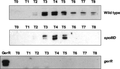
FIG. 4.
Western blot analysis performed with an anti-GerR antibody on extracts of cells collected at various times after the onset of sporulation. The GerR lane of the bottom panel was loaded with proteins extracted 4 h after the onset of sporulation (T4) from a wild-type strain. In all experiments, 25-μg samples of total proteins were fractionated on 15% polyacrylamide gels, electrotransferred to nitrocellulose membranes, reacted first with primary antibodies and then with peroxidase-conjugated secondary antibodies, and visualized by the enhanced chemiluminescence method.

