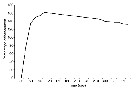
Figure 4b:
Findings in right breast of 43-year-old woman with recent diagnosis of left-sided breast cancer. (a) Axial contrast-enhanced 3D T1-weighted high-spatial-resolution subtraction MR image (7.08/3.56, 10° flip angle) of right breast shows large (11 × 5 × 5-cm) spiculated enhancing mass lesion (arrow). (b) Kinetic curve for same lesion was categorized as type III (washout). (c) Axial ADC map (b values, 0 and 600 sec/mm2; 9548/64; 90° flip angle) shows same lesion (arrow) with restricted diffusion. Absolute ADC of lesion was 1.3 × 10⁻³ mm2/sec, and glandular tissue–normalized ADC was 0.45 × 10⁻³ mm2/sec. Final histopathologic diagnosis was infiltrating ductal carcinoma with areas of ductal carcinoma in situ.

