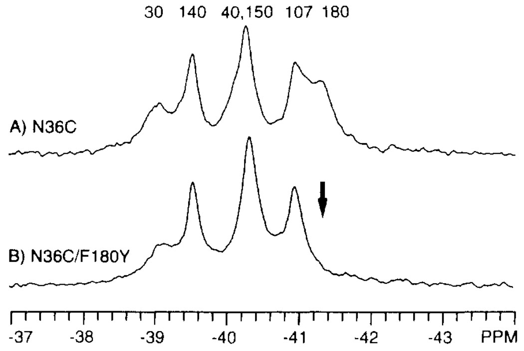
FIGURE 3.
Assignment of 4-F-Phe 19F NMR resonances by site-directed mutagenesis. Shown are the 19F NMR spectra of two 4-F-Phe-labeled domains: N36C (A) and N36C/F180Y (B). The arrow indicates the resonance deleted by the mutation. The final assignments provided by replacement and nudge mutational analysis are indicated (see text). Spectra were obtained at 470 MHz and 25°C in the same buffer as in Figure 2, with the addition of 10% D2O and 50 µM 5-F-Trp. The concentration of dimeric domain was 0.6 mM.

