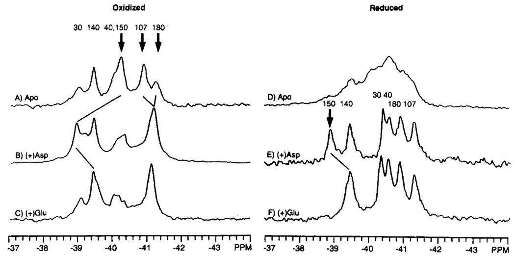
FIGURE 4.
Effect of attractant ligands on the 19F NMR spectrum of the 4-F-Phe labeled ligand binding domain. Shown are the spectra of the apo (A, D), aspartate bound (B, E), and glutamate-bound (C, F) states of the oxidized and reduced N36C ligand binding domain. The bold arrows indicate which of the assigned resonances are shifted by attractant binding; the new positions of these resonances are indicated by the light diagonal lines. Sample conditions were as in Figure 3; where indicated, 5 mM aspartate or 25 mM glutamate was also present. The concentration of dimeric domain was 0.7–2 mM.

