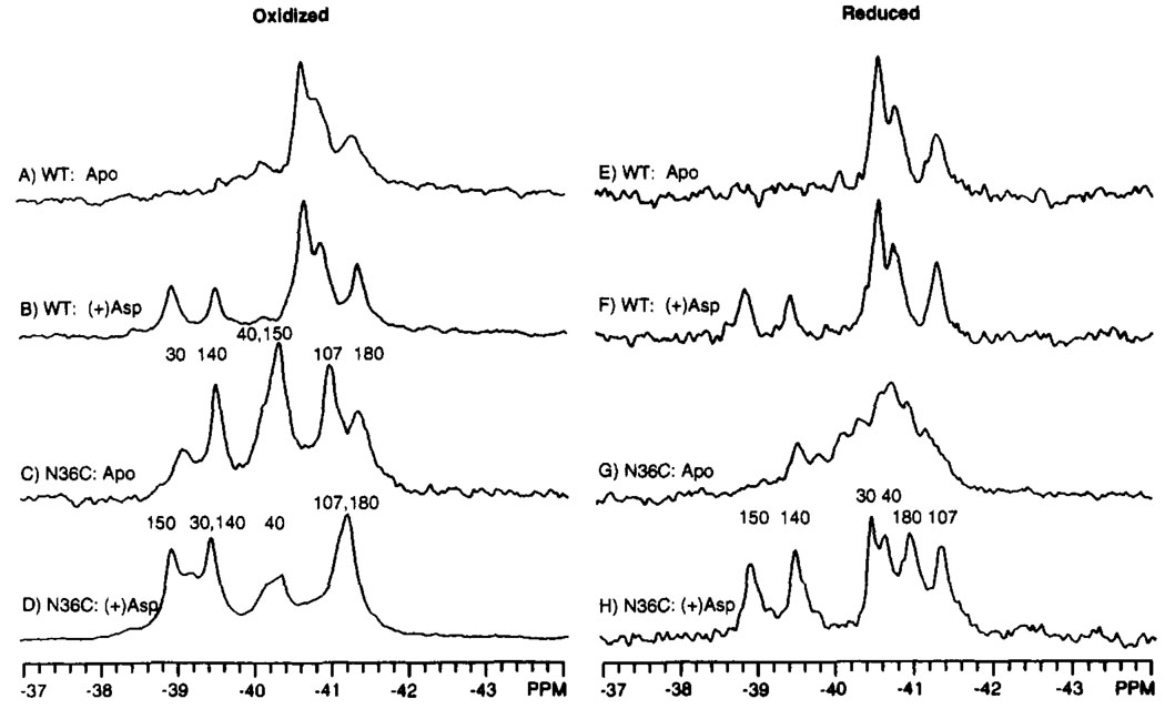
FIGURE 7.
Effect of the Cys36–Cys36′ disulfide on the 19F NMR spectrum of the 4-F-Phe-labeled ligand binding domain. Shown are the spectra of domains under oxidizing (A–D) or reducing (E–H, 50 mM DTT) conditions for both the wild-type and N36C mutant domains. The known assignments are indicated. Sample parameters were as in Figure 3; also present was 5 mM aspartate, where indicated. The concentration of dimeric domain was 0.4–1.0 mM.

