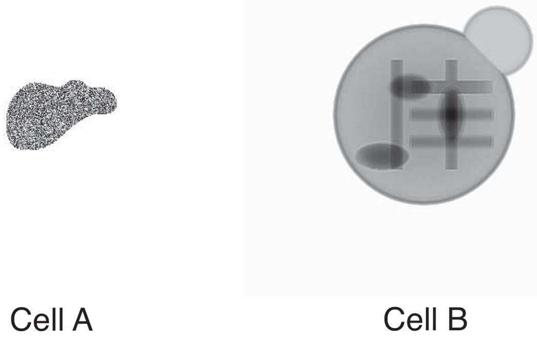
Fig. 2.
Defined objects used for image simulations. Shown here are the magnitudes of the simulated exit waves resulting from plane wave illumination of the objects. Cell A has random protein thicknesses within an irregular boundary, while Cell B has a lipid membrane and several protein bars and ellipses inside.

