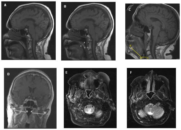
Figure 1.
MRI measurements: (A) Palatal length on sagittal T1-weighted image. (B) Palatal thickness (A-B) and retropalatal distance (C-D) on a sagittal T1-weighted image. (C) Tongue length (A-B) and the retroglossal space (C-D) on sagittal T1-weighted image. (D) Lateral pharyngeal wall thickness on a coronal T1-weighted image. (E) Cross sectional area of high retropharyngeal region on axial T2-weighted image. (F) Cross-sectional area of nasopharynx on axial T2-weighted image.

