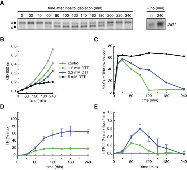
Figure 1. Transient Ire1 activation in non-lethal ER stress conditions.
(A) After depletion of inositol from the growth media, wild type yeast cells were sampled from a master culture every 20 min, and total RNA was purified and subjected to Northern blot analysis using a probe for the first exon of HAC1 mRNA. After a lag phase, HAC1 mRNA splicing displayed activation and deactivation phases. u, unspliced HAC1 mRNA; s, spliced HAC1 mRNA. Right panel: wild type cells 0 min and 240 min after inositol depletion and probed for the INO1 mRNA. (B) Cell growth was monitored over time in wild type cells treated with 5 mM, 2.2 mM, 1.5 mM, and 0 mM DTT by measuring the OD600. Cells treated with 5 mM DTT cease to divide, while cells treated with 2.2 mM or 1.5 mM DTT continue to grow. (C) Wild type cells were treated with 5 mM, 2.2 mM, or 1.5 mM DTT and sampled over time. After Northern blot analysis, the percentage of spliced HAC1 mRNA was quantified (blots are shown in the supplement). Cells treated with 5 mM DTT displayed sustained maximal splicing, while cells treated with 2.2 mM or 1.5 mM displayed transient HAC1 mRNA splicing: the same activation and deactivation phases as the response to the depletion of inositol. (D) Wild type cells were constructed bearing a transcriptional reporter (TR) consisting of four repeats of a UPR-responsive DNA element controlling the expression of GFP. These cells were treated with 2.2 mM, 1.5 mM, or 0 mM DTT, sampled over time, and subjected to flow cytometry to quantify the GFP fluorescence. The TR was induced to dose-dependent plateaus due to the >8 h half life of GFP. % max is defined as the GFP fluorescence in cells treated with 5 mM DTT for 4 h. (E) When plotted as the rate of GFP produced per minute, the TR displayed the same activation and deactivation phases as spliced HAC1 mRNA. Transient Ire1 activation leads to transient transcriptional activation. % max as defined in (D).

