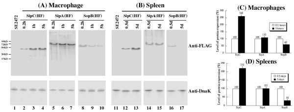
Figure 6.
Western blot analyses of the expression of the tagged proteins from bacterial strains SE2472 (lanes 1 and 11), SipC(HF) (lanes 2-4, 12-13), SipA(HF) (lanes 5-7, 14-15), and SopB(HF)(lanes 8-10, 16-17). In (A), bacterial protein samples were isolated from macrophages at 0.2, 1, and 5 hours of postinfection. In (B), BALB/c mice were intraperitoneally infected with 1 × 10⁶ and 1 × 10⁴ CFU of the tagged strains, and internalized bacteria were recovered from the spleen at 0.5 days and 4 days post inoculation, respectively. The expression of bacterial DnaK was used as the internal control. Protein samples were reacted with antibodies against the FLAG sequence (top panel) and DnaK (low panel). Each lane was loaded with material from 5 × 10⁷ CFU bacteria. (C-D). Level of tagged proteins from the bacterial strains recovered from the macrophages and spleens of infected mice as determined in (A) and (B). The values, which are the means of triplicate experiments, represent the relative percentage of the levels of the tagged proteins in the bacteria recovered from macrophages (C) at 5 hours postinfection and from the spleen at 5 days postinoculation (D), as compared to those in the bacteria recovered from macrophages at 0.2 hours postinfection and from spleen at 0.5 days post inoculation, respectively.

