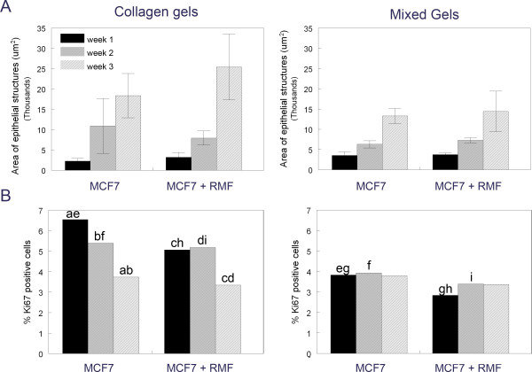
Figure 3.
Morphometric analysis of area and proliferation index of epithelial structures in type I collagen and mixed gels. Panel A: The area was measured as μm2. Panel B: Proliferating cells are expressed as % Ki67 positive cells compared to all cells. Same letters denote significant differences between conditions and time points (p < 0.05).

