
Figure 4.
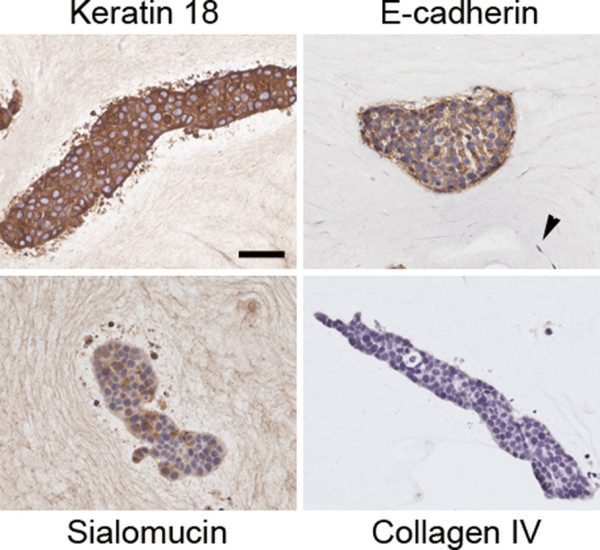
Characterization of epithelial structures in collagen gels after 2 weeks in culture. Co-cultures of MCF7 cells and RMF were characterized using immunohistochemical analyses for luminal epithelial cell origin (Keratin 18), adherens junctions (E-cadherin), and polarization (Sialomucin). No staining was observed for the basement membrane marker type IV collagen. Arrowhead points to fibroblast. Scale bar: 50 μm.