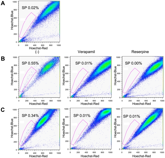
Figure 1. SP cells in high metastatic M3a2 and M4e cells.
(A): Flow cytometry analysis of 686LN cells. Cells were stained with Hoechst 33342. (B and C): M3a2, and M4e cells were stained with Hoechst 33342 in the absence or presence of either verapamil or reserpine. The SP cells were gated and shown as percentage as indicated. Flow cytometry analyses were performed in triplicate with similar results in each case.

