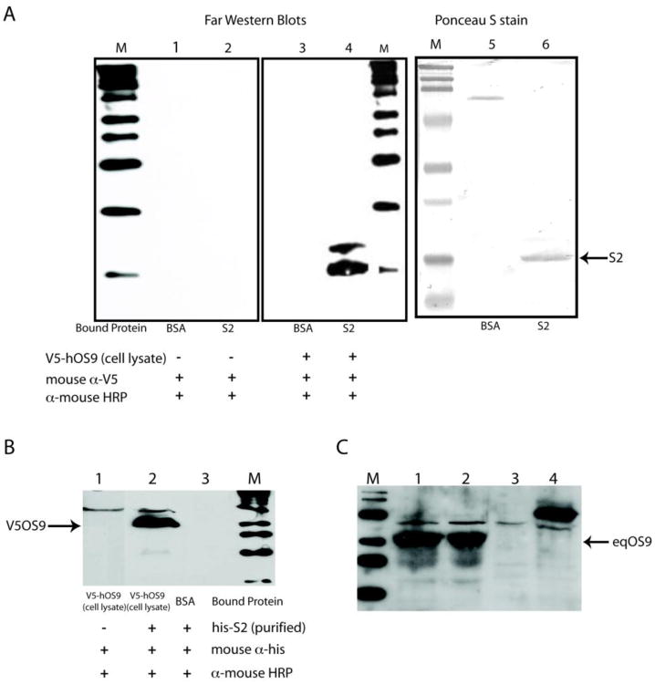
Figure 2.
Panel A. Far western blots showing the interaction of immobilized recombinant S2 with recombinant V5-tagged hOS9 in CHO cell lysates. Lanes 1–3 are reagent controls. Lane 4 shows the binding of V5-OS9 to the membrane at the position of bacterially expressed, histidine tagged S2 (his-S2). The larger product in lane 4 is not present in the controls and may result from V5-OS9 binding to a slower migrating form of his-S2 or alternatively, interaction of V5-OS9 with a contaminating bacterial protein in the lysate. V5-OS9 is detected using mouse anti-V5 antibody. Lanes 5 and 6 show the positions of S2 and the control protein, bovine serum albumin. Panel B. Far western blot showing the interaction of immobilized V5-OS9 with soluble his-S2. Panel C. Western blot using anti-human OS9 indicates that equine macrophages express OS9. Lanes 1 and 2 contain lysates from equine monocyte-derived macrophages. Lane 3 contains a CHO cell lysate. Lane 4 contains a cell lysate from V5-tagged human OS9 transfected CHO cells.

