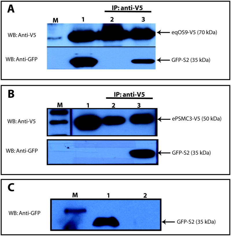
Figure 3.
Co-immunoprecipitation of EIAV S2 with equine OS9 and PMSC3. Panel A. Co-IP of equine OS9-V5 and GFP-S2. Lane 1: Mixed lysates from equine OS9-V5 and S2-GFP transfected cells. Lane 2: Mixed lysates, equine OS9-V5 and GFP-control tranfected cells. Lane 3: Mixed lysates, equine OS9-V5 and GFP-S2 transfected cells. Lanes 2 and 3, immunoprecipitates with anti-V5. Western blots with anti-GFP or anti-V5 as indicated. Panel B. Co-IP of equine PSMC3-V5 and GFP-S2. Lane 1: Lysate from equine PSMC3-V5 transfected cells. Lane 2: ePSMC3-V5 transfected cell lysate mixed with GFP-control lysate; immunoprecipitated with anti-V5. Lane 3: ePSMC3-V5 transfected cell lysate mixed with GFP-S2 transfected cell lysate; immunoprecipitated with anti-V5. Panel C. GFP-S2 control. Lysate from GFP-S2 transfected cells was immunoprecipitated with anti-V5. Lane 1: Column flow-through. Lane 2. Column eluant. In all panels, M designates lanes with protein molecular weight markers.

