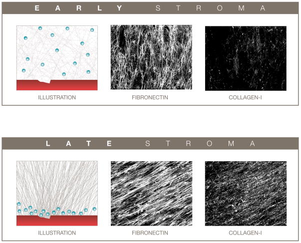
Figure 3.
The penetration of extravasated chemotherapeutic agents from circulation into the tumor microenvironment can be influenced by the tumor-associated stromal compartment. A normal-like less organized and loosely packed early tumor microenvironment (top) can provide for greater drug penetration, while a more organized and tighter packed late tumor microenvironment (bottom) may result in decreased drug penetration. Left panels are representative illustrations of damaged blood vessels (red) and putative extravasated drugs (e.g., DDS, blue spheres) penetrating through the assorted ECMs. Middle and right panels depict reconstructed confocal images of indirect immunofluorescence showing in vivo-like ECM fibronectin (middle) and collagen I (right) fibers derived from assorted fibroblasts. Confocal images are reproductions from Amatangelo, Bassi et al. 2005 [69] presented with permission from the American Journal of Pathology.

