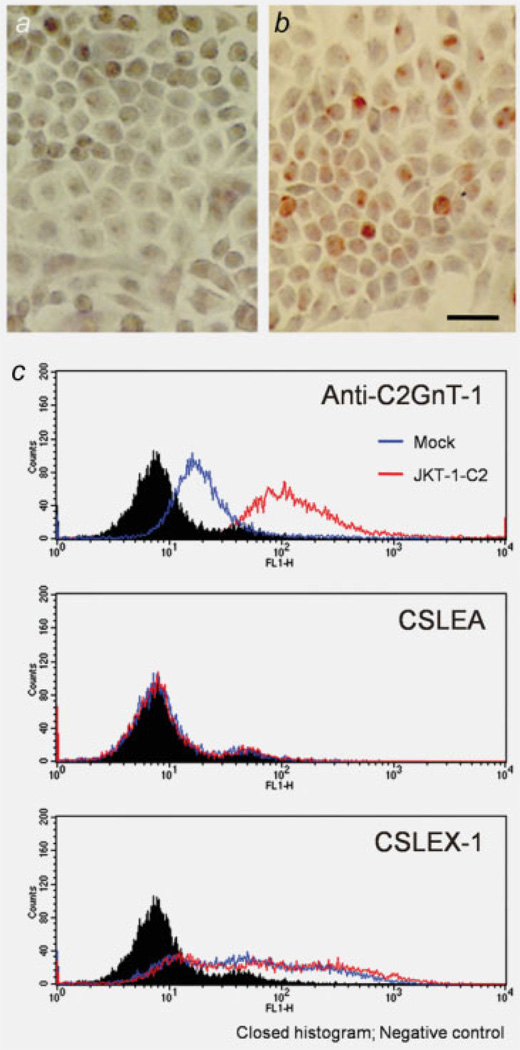
Figure 3.
Immunocytochemical and flow cytometric analyses of JKT-1 cells. On immunocytochemistry using anti-C2GnT-1, the parent JKT-1 cells did not express C2GnT-1 (a), CSLEA (c, middle) but positive in CSLEX-1 (c, bottom). After transfection of mammalian expression vector harboring C2GnT-1 cDNA, these transfected cells became positive for C2GnT-1 (b). C2GnT-1 was also detected in JKT-1-C2 by flowcytometric analysis. Transfection of C2GnT-1 did not influence to the expression pattern of sialyl Lewis A and X (c, middle, bottom). Closed histograms were negative control, open histograms of blue and red were JKT-1-mock and JKT-1-C2 cells, respectively. Scale bar: 10 µm.

