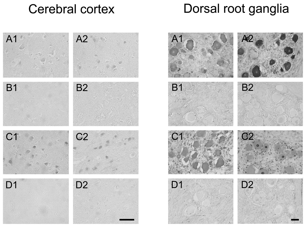
Figure 3.
Immunohistochemical analysis of serum antibody reactivity towards cells in the brain cerebral cortex (left panel) and DRG (right panel). A) Staining of sections with serum from borrelial seropositive (A1) and borrelial seronegative (A2) patients with anti-neural antibody reactivity (as determined by immunoblotting) showed specific binding to neurons of the cerebral cortex and the DRG. B) Staining of sections with serum from borrelial seropositive (B1) and borrelial seronegative (B2) post-Lyme healthy individuals with anti-neural antibody reactivity showed faint or no specific binding of antibodies to cerebral cortex and DRG tissues. C) Serum antibodies from two representative SLE patients (C1 and C2) with anti-neural antibody reactivity bound strongly to neurons and glial cells in the cerebral cortex and the DRG. D) Sera from two normal healthy subjects (D1 and D2) did not stain tissues specifically. Bars = 50 µm.

