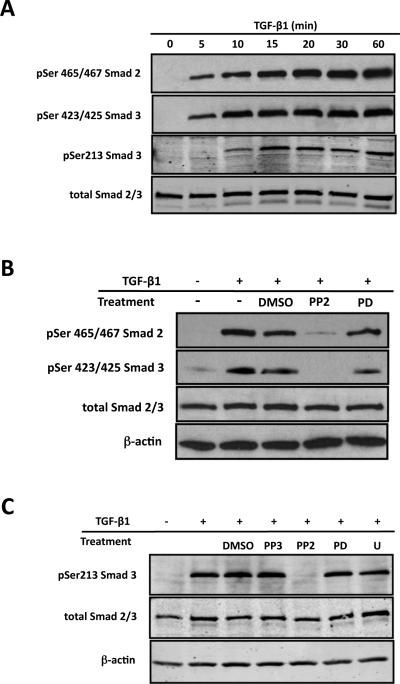
Figure 5. Src regulates Smad activation in an Erk independent fashion.
ROS osteoblast-like cells were cultured until they were 80% confluent and serum starved for 24 hours prior to any treatment. After treatment, whole-cell protein extracts were analyzed by Western blot analysis. Experiment were repeated 4 times with similar results. (A) To determine the time course for TGF-β1 induced Smad2 and Smad3 activation, cells were treated with TGF-β1 (5ng/ml) for 0, 5, 10, 15, 30 or 60 minutes. Both Smad2 and Smad3 demonstrate the robust activation following TGF-β1 treatment with maximal activation occurring 20 minutes post-treatment (B) Serum-starved cells were pre-treated with the Src kinase inhibitor, PP2 (20mM), the Erk inhibitor, PD98059 (PD; 20mM), or diluent (DMSO) alone for 30 minutes followed by treatment with TGF-b1 (5ng/ml) for 20 minutes. While Src inhibition blocks activation of Smad2 and Smad3, Erk inhibition has no effect on Smad activation. (C) Serum-deprived cells were pre-treated with PP2 (20mM; Src kinase inhibitor), PD98059 (PD; 20mM; Erk inhibitor), U0126 (U; 30mM; Erk inhibitor), PP3 (an inactive analog of PP2), or DMSO (diluent only) for 30 minutes followed by TGF-b1 stimulation (5ng/ml) for 20 minutes. The results demonstrated that inhibition of Src, but not Erk, blocks linker region phosphorylation of Smad3.

